
Generative AI is poised to change how chest X-rays are interpreted. Instead of just flagging abnormalities, these models can produce complete radiology reports from the images themselves.
At RSNA 2025, Robert Harris of vRad is developing a generative chest X-ray model. vRad interprets about 7 million studies each year, which provides a massive dataset for training algorithms. This scale allows the model to learn from rare pathologies, improving its accuracy and reliability. The goal is to move beyond simple detection to full report generation, marking a shift in generative AI radiology. This vRad AI model aims to bring practical, actionable insights directly from the image, making chest X-ray report generation more efficient for radiologists.
How Generative AI Creates Chest X-Ray Reports from Images
Generative models take a fundamentally different approach to report generation. Instead of simply filling in predefined templates, these systems learn directly from the way radiologists actually describe what they see. The key training material comes from radiologists’ dictated reports, which serve as the ground truth. By studying thousands of these real-world examples, the model learns the relationship between visual patterns in a chest X-ray and the corresponding medical language.

From Image to Report: The Generative Workflow
When you feed a chest X-ray into a generative model, it doesn’t just pick from a set of canned phrases. Instead, it processes the entire image and produces a full, structured report that includes both the findings and the impression. This is where natural language processing radiology techniques come into play. The model needs to understand not just what’s in the image, but how to describe it in a clinical context—using the right terminology, ordering, and level of detail.
However, getting to this point requires significant upfront work. Training a generative chest xray model demands extensive preprocessing to remove report inconsistencies. Radiologists may use different phrasing for the same condition, or vary how they structure their findings. The training pipeline must normalize these variations so the model learns consistent patterns rather than memorizing noise. This preprocessing step is what makes the difference between a model that produces reliable, coherent reports and one that outputs confusing or contradictory text.
For the radiologist, the result is a report generation AI that feels more like a capable assistant. It can handle the bulk of the descriptive work, leaving you to focus on the clinical judgment and any subtle findings that require human expertise.
Why Chest X-Rays Are the Ideal Starting Point for Generative AI
That kind of reliable assistant doesn’t appear overnight. Getting a generative model to produce accurate, clinically useful text requires a massive amount of training. And the type of data you feed it matters enormously. This is where chest X-rays have a clear advantage over other medical imaging. A single chest X-ray contains about 100 times less data than a CT scan. That might sound like a limitation, but for training a generative AI, it’s actually a major benefit.
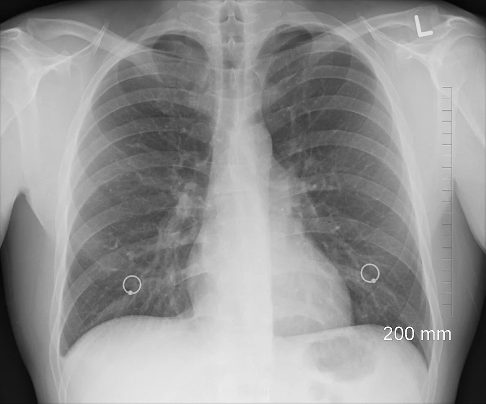
When you compare chest X-ray vs CT data size, the difference is stark. A CT scan builds a 3D picture from many individual slices, creating a huge file. A chest X-ray is a single 2D image. This lower data burden means a generative model can train much faster and requires far fewer computational resources. You don’t need a supercomputer to run the training pipeline; a powerful workstation can often handle the job. This makes the technology more accessible for research hospitals and smaller clinics that might not have access to massive cloud computing budgets.
The simpler structure of a chest X-ray also helps with a common problem in generative AI: hallucinations. With less complex data to interpret, the model has fewer opportunities to invent false details. It learns the clear patterns of anatomy and pathology without getting lost in the noise of hundreds of thin slices. This focus on generative model efficiency means you get a tool that is not only faster to train, but also more reliable in its output right from the start.
Training Challenges: Report Inconsistencies and Rare Pathologies
That efficiency is critical, but it doesn’t solve every problem in building a reliable generative chest xray model. The raw data itself — the radiology reports — presents a major hurdle. Reports vary wildly in style and completeness. One radiologist might write “no acute cardiopulmonary abnormality,” while another describes the same finding as “clear lungs and normal cardiac silhouette.” This variation means training requires extensive preprocessing to remove report inconsistencies. Without that cleanup, the model learns to associate different phrases with the same condition, which leads to confusion and unreliable image generation.
What Inconsistencies Are Removed During Preprocessing?
You might wonder what exactly needs to be scrubbed. Common issues include variable terminology, missing sections like “impression” or “findings,” and differing levels of detail — some reports are terse, others paragraph-length. Preprocessing involves radiology report normalization: standardizing terms, filling in missing structure, and trimming excess text. This step ensures the model sees consistent language for each pathology, which directly improves its ability to generate accurate chest X-rays.
Then there is the challenge of rare pathologies. Many AI models struggle because they simply do not see enough examples of uncommon conditions during training. For a generative chest xray model to handle cases like a superior mesenteric artery occlusion, it needs exposure to that specific finding. This is where a large dataset matters. vRad interprets 7 million studies annually, enabling development of algorithms for rare pathologies. That volume means the model can encounter and learn from these edge cases, making it far more practical for real-world use. Without such a broad data pool, rare disease AI training would remain a theoretical exercise rather than a clinical reality.
vRad’s Investigational Generative Model: Current Status and Future Plans
This wealth of clinical data, rich with diverse cases, provides the perfect foundation for vRad’s own AI development efforts. Still, their investigational generative chest X-ray model isn’t quite ready for clinical deployment. It remains under active study as part of a careful validation process, ensuring everything works as intended before it reaches patients.

Why vRad Builds Its Own AI Models
Why does vRad choose to build its own tools instead of relying solely on third-party vendors? The key reason is their extensive quality assurance program. This program generates a massive, high-quality dataset uniquely suited for training specialized AI models. By developing AI in-house, vRad can train the model directly on the specific data and workflows its radiologists use every day. This investigational phase is critical for confirming safety and accuracy before any clinical rollout.
As for when you might see this generative chest X-ray technology in active clinical use, the exact AI deployment timeline hasn’t been announced yet. The company is prioritizing a methodical approach over speed. This means the investigational AI radiology model will undergo rigorous testing to meet the high standards demanded in medicine. For now, the focus remains on the study, gathering the evidence needed to prove the model’s real-world value.
Accuracy, Incidental Findings, and Regulatory Path for Generative Chest X-Ray AI
As that evidence gathering continues, one of the most promising aspects of the generative approach is its potential to address a persistent challenge in radiology: incidental findings. These are abnormalities visible on an image that fall outside the original clinical question — for example, spotting a lung nodule while looking for pneumonia. Research shows that many radiology misses are precisely these incidental findings, simply because the radiologist’s focus was directed elsewhere. A generative chest xray model could help by automatically including all visible abnormalities in its output, reducing the chance that something important gets overlooked.
How Does the Model Handle Incidental Findings?
The traditional approach to AI in radiology has been single-pathology models — one tool for pneumonia, another for nodules, a third for fractures. Each operates in isolation. A generative model, by contrast, creates a full report describing everything it sees on the image. In theory, this means it flags findings the original exam wasn’t looking for. The practical benefit is clear: a report that mentions an unexpected finding gives the referring physician a chance to follow up, where a silent miss would not. However, the accuracy of this approach compared to traditional single-pathology AI is still being studied. Early results are promising, but it’s too soon to say whether generative models outperform specialized tools for every type of finding.
Ensuring Report Completeness and Avoiding Hallucinations
One concern with any generative AI is hallucination — the model describing something that isn’t actually there. In radiology, a false positive could trigger unnecessary follow-up scans or procedures. Developers are working on safeguards, such as training the model to only describe features present in the image and cross-referencing its output with known anatomical boundaries. The goal is report completeness without misleading additions. Since the technology is still investigational in many practices, these guardrails are being tested alongside the model itself, not after deployment.
Before any generative radiology AI reaches widespread clinical use, it will need regulatory clearance — typically from bodies like the FDA. That process requires demonstrating both safety and effectiveness across diverse patient populations and imaging settings. Accuracy data must show the model performs consistently, not just on clean study datasets but on real-world scans with varying quality. The path is rigorous by design, and while it slows adoption, it also builds the trust necessary for radiologists and patients to rely on the technology. For now, the generative chest xray approach remains under evaluation, but its potential to catch incidental findings and improve report completeness makes it one to watch.
Frequently Asked Questions
How does generative AI write a chest X-ray report from the image alone?
Generative chest xray models analyze the pixel data in an X-ray image to identify patterns linked to specific findings, like opacities or cardiomegaly. The AI then drafts a structured report in natural language, summarizing these observations. You typically still have a radiologist review and finalize the report for accuracy.
Why are chest X-rays the starting point for generative AI instead of CT scans?
Chest X-rays are more common, faster to acquire, and produce simpler 2D images compared to the complex 3D data from CT scans. This makes them a practical and lightweight dataset for training generative models. The high volume of routine chest X-rays also provides the large, standardized image sets needed for reliable model development.
What challenges does training generative AI models face with radiology reports?
A key challenge is the variability in how radiologists write reports, including different terminology and levels of detail. The AI must learn to associate image features accurately with often-subjective textual descriptions. Another practical hurdle is ensuring patient data privacy during the training process, which requires careful de-identification steps.