Magnetic resonance imaging tech is a non-invasive imaging technology that produces detailed three-dimensional anatomical images without using ionizing radiation. It works by aligning protons with a strong magnetic field and pulsing radiofrequency current. As protons realign, sensors detect energy release, enabling tissue differentiation. Magnetic resonance imaging (MRI) is a non-invasive imaging technology that uses magnetic fields and radio waves to produce three-dimensional detailed anatomical images. This guide will explore how MRI technology functions, its diverse clinical applications, crucial safety considerations, and the innovative advancements being driven by the National Institute of Biomedical Imaging and Bioengineering (NIBIB).
What Is Magnetic Resonance Imaging Tech and How Does It Work?
Magnetic resonance imaging (MRI) is a non-invasive imaging technology that produces three-dimensional detailed anatomical images. This advanced diagnostic tool operates using powerful magnetic fields and radio waves to generate highly detailed visuals of the body’s internal structures. Unlike technologies that use X-rays, MRI is based on technology that excites and detects changes in the rotational axis of protons in water within living tissues. The process begins when MRIs employ powerful magnets that produce a strong magnetic field, which aligns the protons in the body.
When a radiofrequency current is pulsed through the patient, protons are stimulated and spin out of equilibrium. As these protons are nudged out of alignment, they absorb energy, and when the radiofrequency field is turned off, the MRI sensors detect the energy released as protons realign with the magnetic field. This realignment process varies; the time for protons to realign and the amount of energy released depend on the specific environment and the chemical nature of the molecules they are part of.
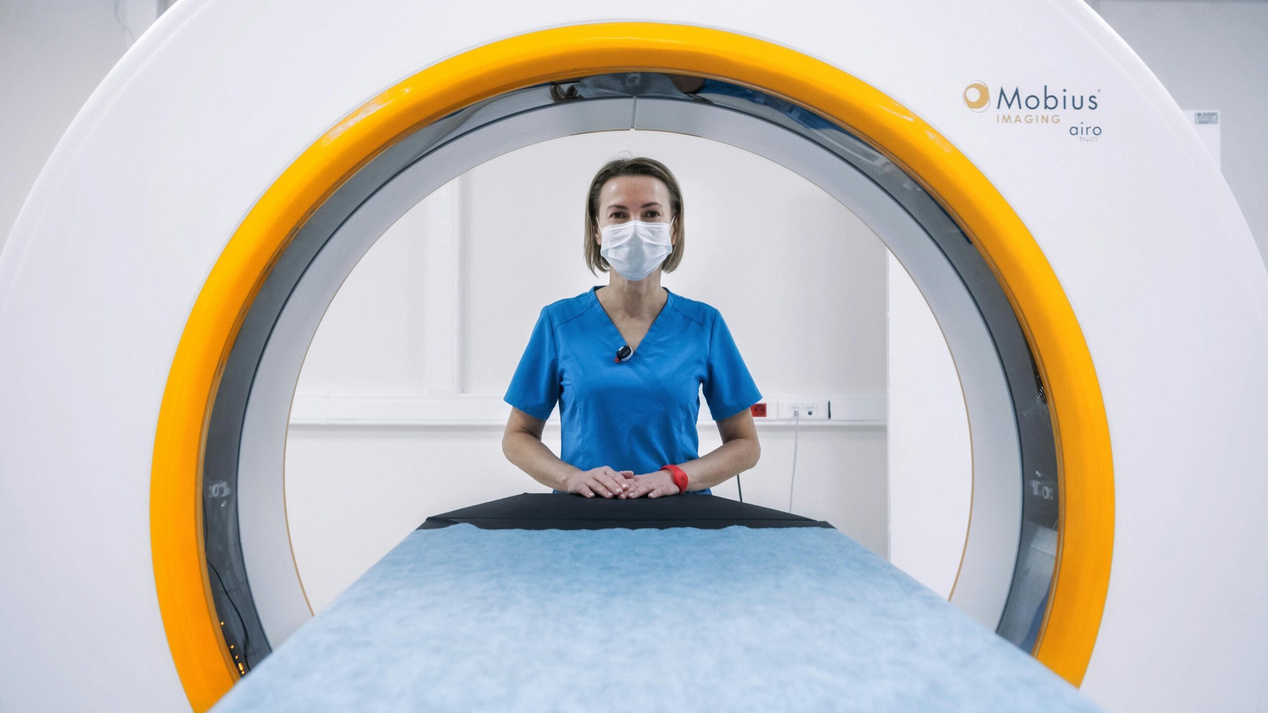
Physicians can differentiate between tissue types based on these subtle variations in magnetic properties. To obtain an MRI image, a patient is placed inside a large magnet and must remain still during the entire imaging process to ensure clear and accurate results. Crucially, MRI does not use damaging ionizing radiation of X-rays, unlike computed tomography (CT). This makes it a safer option for patients requiring repeated imaging or those who are particularly sensitive to radiation exposure.
Let’s explore why these sophisticated mechanisms matter in clinical practice.
What Are the Clinical Applications of MRI?
MRI is often used for disease detection, diagnosis, and treatment monitoring across a wide spectrum of medical conditions. Its ability to produce exceptionally detailed images of soft tissues makes it invaluable in modern medicine. MRI scanners are particularly well-suited to image non-bony parts or soft tissues of the body, offering clarity that other imaging modalities often cannot match.
The brain, spinal cord, nerves, muscles, ligaments, and tendons are seen more clearly with Magnetic Resonance Imaging (MRI) than with X-rays and CT scans. This precision is critical for diagnosing conditions ranging from neurological disorders and sports injuries to internal organ abnormalities and certain types of cancer. Furthermore, a specialized technique called Functional Magnetic Resonance Imaging (fMRI) is a specialized MRI used to observe brain structures and determine which areas activate during cognitive tasks. This allows researchers and clinicians to map brain activity in real-time, aiding in the understanding of conditions like stroke, epilepsy, and brain tumors.
What can MRI show that CT cannot? MRI excels in visualizing soft tissues with superior contrast resolution compared to CT scans, particularly for brain, spinal cord, and musculoskeletal imaging, without the use of ionizing radiation.
Understanding these applications naturally leads to questions about safety.
Is MRI Safe? Understanding Risks and Precautions
Magnetic Resonance Imaging (MRI) is generally considered a safe imaging technique, largely due to its lack of ionizing radiation. However, the powerful magnetic fields used necessitate strict safety protocols. MRI exerts powerful forces on magnetizable objects, strong enough to fling a wheelchair across the room if it enters the magnetic field improperly.
Therefore, people with implants containing iron should not enter an MRI machine. This includes a wide range of devices such as pacemakers, vagus nerve stimulators, implantable cardioverter-defibrillators, loop recorders, insulin pumps, cochlear implants, deep brain stimulators, and even capsules from capsule endoscopy. It is critical for patients to disclose all medical implants and devices before an MRI scan. The loud noise during MRI scanning can reach 120 decibels, comparable to a rock concert, and may require ear protection for patients to prevent hearing damage.
Contrast agents containing Gadolinium may be given intravenously before or during MRI to increase proton realignment speed, enhancing the visibility of certain tissues and abnormalities. However, patients with severe renal failure requiring dialysis may risk nephrogenic systemic fibrosis, a rare but serious condition linked to certain gadolinium-containing agents like gadodiamide. Current guidelines in the United States recommend that dialysis patients receive gadolinium agents only when essential, with prompt dialysis performed after the scan to help clear the agent from the body. No effects of MRI on the fetus have been demonstrated, but MRI is recommended to be avoided as a precaution, especially in the first trimester of pregnancy, unless the benefits clearly outweigh the potential risks.
Safety concerns can be managed with proper precautions and patient comfort approaches.
How Does Functional MRI (fMRI) Work?
Functional Magnetic Resonance Imaging (fMRI) is a specialized MRI technique used to observe brain structures and determine which areas activate during cognitive tasks. It offers a dynamic view of brain function by detecting changes in blood flow, which are indirectly related to neural activity. When a specific area of the brain becomes more active, it requires more oxygen, leading to an increase in blood flow to that region.
fMRI capitalizes on the magnetic properties of oxygenated and deoxygenated hemoglobin. As blood flow increases to active brain regions, the ratio of oxygenated to deoxygenated hemoglobin changes. The MRI scanner detects these subtle changes, generating maps that highlight which parts of the brain are engaged during specific tasks, such as reading, problem-solving, or even responding to stimuli. This capability makes fMRI invaluable for neurological research, understanding brain disorders, and in some cases, for pre-surgical planning.
fMRI is just one of many NIBIB-funded innovations in MRI.
What Innovations in MRI Are Funded by NIBIB?
The National Institute of Biomedical Imaging and Bioengineering (NIBIB) actively funds research to advance MRI technology, improving its diagnostic capabilities and patient experience. In the realm of liver disease, where chronic liver disease and cirrhosis affect millions in the United States, NIBIB-funded researchers developed Magnetic Resonance Elastography (MRE). This innovative method transforms sound waves into images that precisely map the stiffness of the liver. MRE is positioned as a safer, more comfortable, and less expensive alternative to a traditional liver biopsy, with the potential to detect cancer by recognizing slight differences in tissue density.
Furthermore, the National Institute of Biomedical Imaging and Bioengineering is funding crucial research aimed at optimizing MRI for pediatric patients. This includes the development of a robust pediatric body MRI protocol using a specialized pediatric coil designed for smaller bodies. This innovation aims to improve image quality and potentially reduce the need for anesthesia in children undergoing MRI scans. Addressing motion artifacts, a common challenge in MRI, a National Institute of Biomedical Imaging and Bioengineering-funded researcher is developing a sophisticated motion correction system. This system uses optical tracking to adapt MRI pulses to patient pose changes in real time, ensuring clearer images even if the patient moves slightly.
In a significant breakthrough for prostate cancer diagnosis, National Institute of Biomedical Imaging and Bioengineering-funded researchers discovered a way to inject hyperpolarized carbon 13 into prostate cancer patients. This technique allows for the measurement of tumor metabolic rate, providing a fast and accurate picture of tumor aggressiveness and improving risk prediction for prostate cancer patients. Data shows these diverse innovations, funded by NIBIB, highlight the institute’s commitment to making MRI more effective, accessible, and informative.
These innovations also address patient comfort, especially for those with claustrophobia.
What Are the Options for Patients with Claustrophobia?
For patients experiencing claustrophobia, several coping mechanisms and technological advancements offer solutions to undergo Magnetic Resonance Imaging (MRI) comfortably. These strategies aim to mitigate anxiety associated with the confined space of traditional MRI scanners. Coping mechanisms include familiarization with the scanner environment, visualization techniques to mentally escape the tight space, and in some cases, the use of sedation or even anesthesia for severe cases. Patients can also find relief by listening to music, watching a video during the scan, and having access to a panic button that allows them to stop the procedure immediately if needed.
Open MRI technology was specifically developed for patients who are uncomfortable with the narrow tunnel and noise of traditional MRI machines. These scanners have a more open design, with magnets positioned above and below the patient, allowing for scans to be performed without the feeling of enclosure. Open MRI is also beneficial for patients whose size or weight make traditional MRI impractical or uncomfortable.
Looking ahead, NIBIB continues to push the boundaries of MRI technology.
Conclusion: The Future of Magnetic Resonance Imaging Tech
Magnetic resonance imaging tech stands as a cornerstone of modern medical diagnostics, offering detailed anatomical insights without the risks associated with ionizing radiation. MRI does not use damaging ionizing radiation, unlike computed tomography (CT), making it a preferred choice for many diagnostic procedures. Innovations, such as Magnetic Resonance Elastography (MRE), developed with funding from the National Institute of Biomedical Imaging and Bioengineering (NIBIB), are further expanding its capabilities, offering safer and more effective methods for diagnosing conditions like chronic liver disease. Reports indicate NIBIB’s continued investment in MRI research promises even more advanced applications, improving diagnostic accuracy, patient comfort, and ultimately, health outcomes.
FAQ
Q: What is magnetic resonance imaging (MRI)?
A: MRI is a non-invasive imaging technology that uses a strong magnetic field and radio waves to produce detailed three-dimensional anatomical images of the body’s soft tissues, without using ionizing radiation.
Q: Is MRI safe?
A: MRI is generally safe, but it uses powerful magnets that can attract metal objects. People with certain implants (like pacemakers) or severe renal failure may have risks. Noise can reach 120 decibels, so ear protection is often needed.
Q: What is functional MRI used for?
A: Functional MRI (fMRI) is a specialized MRI that measures brain activity by detecting changes in blood flow. It is used to observe brain structures and determine which areas become active during cognitive tasks or in response to stimuli.
Q: What is magnetic resonance elastography (MRE)?
A: MRE is a NIBIB-funded MRI innovation that uses sound waves to create an elasticity map of soft tissues, particularly the liver. It is safer and more comfortable than a liver biopsy and can also help detect cancer by identifying tissue stiffness.
Q: Can I have an MRI if I am pregnant?
A: While no harmful effects on the fetus have been demonstrated, MRI is generally avoided during the first trimester as a precaution. If clinically necessary, it may be performed.
Q: What should I do if I am claustrophobic during an MRI?
A: Options include familiarization with the scanner, sedation, anesthesia, listening to music, watching a video, or using a panic button. Open MRI machines are also available and are less confining.